Background
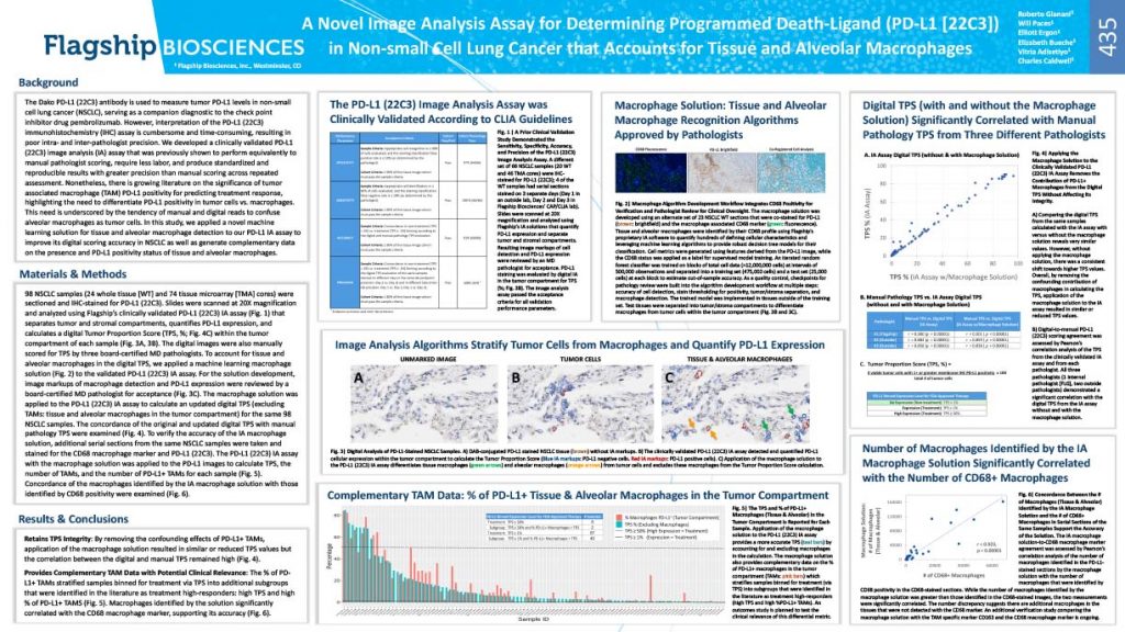
The Dako PD-L1 (22C3) antibody is used to measure tumor PD-L1 levels in non-small cell lung cancer (NSCLC), serving as a companion diagnostic to the check point inhibitor drug pembrolizumab. However, interpretation of the PD-L1 (22C3) immunohistochemistry (IHC) assay is cumbersome and time-consuming, resulting in poor intra- and inter-pathologist precision. We developed a clinically validated PD-L1 (22C3) image analysis (IA) assay that was previously shown to perform equivalently to manual pathologist scoring, require less labor, and produce standardized and reproducible results with greater precision than manual scoring across repeated assessment. Nonetheless, there is growing literature on the significance of tumor associated macrophage (TAM) PD-L1 positivity for predicting treatment response, highlighting the need to differentiate PD-L1 positivity in tumor cells vs. macrophages. This need is underscored by the tendency of manual and digital reads to confuse alveolar macrophages as tumor cells. In this study, we applied a novel machine learning solution for tissue and alveolar macrophage detection to our PD-L1 IA assay to improve its digital scoring accuracy in NSCLC as well as generate complementary data on the presence and PD-L1 positivity status of tissue and alveolar macrophages.
Results and Conclusions
Retains TPS Integrity: By removing the confounding effects of PD-L1+ TAMs, application of the macrophage solution resulted in similar or reduced TPS values but the correlation between the digital and manual TPS remained high (see poster, Fig. 4).
Provides Complementary TAM Data with Potential Clinical Relevance: The % of PD- L1+ TAMs stratified samples binned for treatment via TPS into additional subgroups that were identified in the literature as treatment high-responders: high TPS and high % of PD-L1+ TAMS (see poster, Fig. 5). Macrophages identified by the solution significantly correlated with the CD68 macrophage marker, supporting its accuracy (see poster, Fig. 6).
View more on our work with clinical diagnostics, additional Flagship posters, or contact us for more information.

